DEVELOPMENT OF A SYSTEMATIC APPROACH TO DIAGNOSTIC ULTRASOUND IN NILE TILAPIA Oreochromis niloticus
Ultrasound imaging uses high-frequency sound waves (2 to > 55 MHz) to visualize internal anatomy for diagnostic procedure s. Ultrasonography has been used in aquaculture and fisheries sinc e the early 1980s; however, its widespread use and potential contribution is limited by high variability in equipment, diverse fish morphology, and inadequate reporting of control settings used. The goal of this study was to develop systematic fish handling and ultrasound scanning procedures for viewing internal anatomy and reproductive organs in Nile Tilapia. The objectives were to (1) assess fish handling and probe positioning procedures; (2) manipulate and evaluate pr imary ultrasound control settings (frequency, focus, depth, gain) at three external anatomy landmarks; and, (3) develop systematic, reliable, reproducible fish handling and ultrasound procedures .
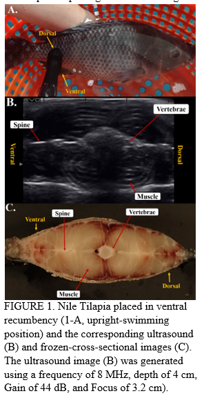
Adult Nile Tilapia (24 females: 24 males) were scanned using the EVO II Ultrasound Scanner (E.I Medical Imaging, CO) equipped with a (6-14 MHz) multiple-frequency range waterproof probe in lateral, ventral, and dorsal recumbence . Probe positioning and settings were developed on external anatomy landmarks (Figure 1). C ross-sectional ultrasound images on the transverse plan (Figure 1-B), and the gross morphology of frozen fish were recorded (Figure 1-C) . Fish positioning, probe placement at external landmarks, control settings, and data obt ained from the frozen cross-section were used to inform image interpretation and development of a systematic ultrasonography approach to generate new data for reproduction research on Nile Tilapia and to create a diagnostic ultrasound user- guide for tilapia hatcheries.
